Bioactive, Human PD-L1 Protein Dimer, His-Avi Tag
| Product Code | CSP-24094-03 |
| Expression Host | HEK293T |
| Verified Applications | ELISA and BLI for PD-L1-specific antibody and BLI for PD-1 ligand protein binding assays. |
| Suggested Applications | SPR for PD-L1-specific antibody and PD-1 protein binding assays. Animal immunization, RUO. |
| Purity | Greater than 90% dimer form as determined by SDS-PAGE under non-reducing condition |
| Amino Acid Range | F19-238R |
For Research Use Only (RUO)
Price: $125.00
Price: $195.00
Price: $350.00
Price: $750.00
Price: $2,500.00
SDS-PAGE
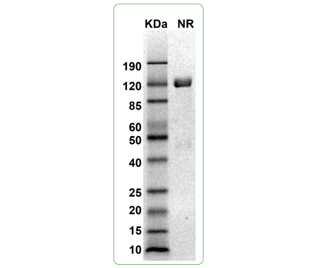
NR: PD-L1 dimer under non-reduced condition
Specifications
Formulation: 0.22μm filtered PBS, pH 7.4
Shipping: Frozen Dry Ice
Storage: -80°C
Human programmed death-ligand 1 (PD-L1), is a type I membrane protein in the immunoglobulin superfamily and a member of the B7 Family of ligands. The recombinant PD-L1 protein dimer (CSP-24094-03) is a cis-homodimer (cis-dimer) and contains a PD-L1 extracellular domain (UniProt# Q9NZQ7, amino acids Phe19-Arg238) fused with a proprietary cis-dimer motif followed by a tandem His-Avi tag at the C-terminus. This dimeric protein is expressed in HEK293T cells. The recombinant human PD-L1 protein dimer is bioactive and can bind to PD-1. It also binds PD-L1-specific antibodies. This PD-L1 protein dimer can be used as an antigen for in vitro assays and antibody screening, and as an immunogen for immunization to generate antibodies targeting more conformational epitopes.
Protein Name: PD-L1
UniProt #: Q9NZQ7
Predicted Molecular Weight: 70 kDa
SDS PAGE Molecular Weight: The migration range of the dimer under non-reducing conditions is 85-120 kDa on SDS PAGE.
Protein Construct: PD-L1 protein dimer contains a PD-L1 extracellular domain (UniProt# Q9NZQ7) fused with a proprietary cis-dimer motif followed by a tandem His-Avi tag at the C-terminus.
Background
Human programmed death-ligand 1 (PD-L1), is a Type I transmembrane protein in the immunoglobulin superfamily and a member of the B7 Family of ligands. PD-L1 is also known as cluster of differentiation 274 (CD274), B7 homolog 1 (B7H1, B7-H1), PDCD1L1, PDCD1LG1, and CD274 molecule. PD-L1 contains an extracellular domain with a distal immunoglobulin V-like (Ig-V-like) domain and proximal immunoglobulin C-like (Ig-C-like) domain, a transmembrane domain, and a cytoplasmic domain. PD-L1 is expressed on T cells, NK cells, macrophages, myeloid DCs, B cells, epithelial cells, and vascular endothelial cells. PD-L1 serves as an immunosuppressive ligand for PD-1 and the overexpression of PD-L1 on many tumor cells can prevent the immune system from attacking tumors. Inhibition of the interaction between PD-1 and PD-L1 can enhance antitumor activity, which has led to a new class of drugs called PD-1 inhibitors to activate the immune system and treat certain types of cancer. PD-L1 is highly expressed in a variety of malignancies, particularly lung cancer. PD-L1 exists as both a monomer and a dimer. Therefore, a recombinant protein mimicking the PD-L1 protein dimer conformation can be crucial for cancer therapeutic discovery.
Alternate Names: cluster of differentiation 274, CD274, B7-H, B7 homolog 1, B7H1, PDCD1L1, PDCD1LG1, PDL1, CD274 molecule, Programmed cell death ligand 1, hPD-L1